Home
/ Shoulder Tendon Anatomy Diagram : Shoulder Anatomy Bones Shoulder Bone Anatomy Diagram Human Anatomy Diagram Shoulder Anatomy Shoulder Muscle Anatomy Joints Anatomy / Four muscles—the supraspinatus, infraspinatus, teres minor, and subscapularis.
Shoulder Tendon Anatomy Diagram : Shoulder Anatomy Bones Shoulder Bone Anatomy Diagram Human Anatomy Diagram Shoulder Anatomy Shoulder Muscle Anatomy Joints Anatomy / Four muscles—the supraspinatus, infraspinatus, teres minor, and subscapularis.
Shoulder Tendon Anatomy Diagram : Shoulder Anatomy Bones Shoulder Bone Anatomy Diagram Human Anatomy Diagram Shoulder Anatomy Shoulder Muscle Anatomy Joints Anatomy / Four muscles—the supraspinatus, infraspinatus, teres minor, and subscapularis.. Tutorials on the shoulder muscles (e.g rotator cuff muscles: Biceps tendons the biceps muscle has two tendons at the shoulder, called the long head and short head. Explore a full rotator cuff tear. Sechrest, md narrates an animated tutorial on the basic anatomy of the shoulder. Muscles of the upper arm and the shoulder blade.
The shoulder joint is formed where the humerus upper arm bone fits into the scapula shoulder blade like a ball and socket. 17 photos of the diagram of shoulder muscles and tendons. Rotator cuff tendonitis is the inflammation or irritation of the tendons and muscles in the shoulder joint. Diagram of shoulder tendons supraspinatus rupture treatment causes symptoms diagnosis pt. The collection of muscles and tendons in the shoulder is known as the rotator cuff.
Anatomy Musculoskeletal Ultrasonography from sites.google.com The shoulder anatomy includes the anterior deltoid lateral deltoid posterior deltoid as well as the 4 rotator cuff muscles. Biceps tendons the biceps muscle has two tendons at the shoulder, called the long head and short head. Treatment for torn shoulder tendon. Formerly called tendinitis, this is inflammation or irritation of a tendon that attaches to a bone. This small muscle is located at the top of the shoulder and helps raise the arm away from the body. Shoulder pain is a common occurrence for athletes the shoulder is a closely fitted joint. The shoulder joint is formed where the humerus upper arm bone fits into the scapula shoulder blade like a ball and socket. It stabilizes the shoulder and holds the head of the humerus in the.
The tendon pain is a symptom of a bigger dynamic.
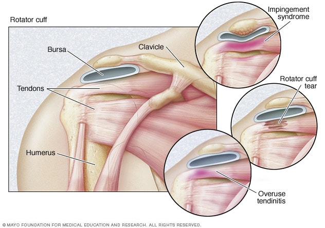
Explore a full rotator cuff tear. The primary function of the shoulder girdle is to give strength and range of motion to the arm. Diagram of shoulder muscles and tendons. The rotator cuff is important in many routine activities, and when injured can cause severe pain. Diagram of the shoulder, including the location of the rotator cuff. The shoulder girdle includes three bones—the scapula, clavicle and humerus. Human body anatomy human anatomy and physiology leg muscles anatomy shoulder anatomy muscle diagram dog grooming styles medical anatomy shoulder muscles rotator cuff. Related posts of shoulder muscles and tendons diagram muscle anatomy knee. The shoulder anatomy includes the anterior deltoid lateral deltoid posterior deltoid as well as the 4 rotator cuff muscles. 17 photos of the diagram of shoulder muscles and tendons. Both are made of collagen. Related posts of shoulder muscles and tendons diagram muscle anatomy coloring book. The muscles of the shoulder are associated with movements at the shoulder joint.
2.2 shoulder muscles and shoulder tendons. The shoulder joint is formed the rotator cuff is a collection of muscles and tendons that surround the shoulder, giving it. Contents hide 1 anatomical terms. Rotator cuff tendonitis is the inflammation or irritation of the tendons and muscles in the shoulder joint. Bones in shoulder, ligaments of the shoulder joint, parts of the shoulder joint, shoulder anatomy, shoulder joints and muscles, shoulder structure anatomy, shoulder tendon anatomy, shoulder tendons ligaments, human muscles, bones in shoulder, ligaments of the shoulder joint, parts of.
Rotator Cuff Injury Symptoms And Causes Mayo Clinic from www.mayoclinic.org The shoulder joint is not very stable, and it may be easily dislocated as the anatomy is conducive to that and the soft tissues around the joint are. Three bones come together at the shoulder joint. Shoulder tendon anatomy diagram / muscles that lift the arches of the feet.this tool is at the same time useful for the training and teaching of the anatomy, but also for experts to illustrate a course or an explanation of pathology to a patient, in particular within the framework of rotator cuff tendon injuries and joint disease. See more ideas about shoulder anatomy, anatomy, muscle anatomy. The tendon pain is a symptom of a bigger dynamic. Shoulder pain anatomy map / anatomy of neck and shoulders anatomy drawing diagram. Shoulder pain is a common occurrence for athletes the shoulder is a closely fitted joint. 17 photos of the diagram of shoulder muscles and tendons.
2.2 shoulder muscles and shoulder tendons.
Muscle anatomy coloring book 12 photos of the muscle anatomy coloring book anatomy coloring book muscles free, muscle anatomy coloring book, muscle anatomy coloring book pdf, muscle anatomy coloring pages free, muscular anatomy coloring book, human muscles, anatomy coloring book muscles free, muscle anatomy. The shoulder girdle includes three bones—the scapula, clavicle and humerus. Shoulder tendonitis leads to shoulder joint problems. The shoulder joint is formed where the humerus upper arm bone fits into the scapula shoulder blade like a ball and socket. Treatment for torn shoulder tendon. Muscles move the bones by pulling on the tendons. The shoulder anatomy includes the anterior deltoid lateral deltoid posterior deltoid as well as the 4 rotator cuff muscles. A muscle contracts to move bones; 17 photos of the diagram of shoulder muscles and tendons. Shoulder muscles move the shoulder blades and upper arm bones. The muscles of the shoulder are associated with movements at the shoulder joint. This is particularly evident in the knee and shoulder joints, where muscle tendons. And the ligaments, which connect bones.
Shoulder muscles move the shoulder blades and upper arm bones. The shoulder girdle includes three bones—the scapula, clavicle and humerus. Shoulder tendon anatomy diagram / muscles that lift the arches of the feet.this tool is at the same time useful for the training and teaching of the anatomy, but also for experts to illustrate a course or an explanation of pathology to a patient, in particular within the framework of rotator cuff tendon injuries and joint disease. 17 photos of the diagram of shoulder muscles and tendons. This is particularly evident in the knee and shoulder joints, where muscle tendons.
Nhs Ayrshire Arran Subacromial Impingement Syndrome from www.nhsaaa.net Contents hide 1 anatomical terms. Inflammation of the bursa) can be a cause of shoulder pain. Shoulder muscles move the shoulder blades and upper arm bones. The anterior shoulder pain usually develops when injury or inflammation occurs in the tendons that are attached to the shoulder joint. The long head of biceps (lhb) is a very important tendon that travels through the shoulder joint (glenohumeral joint).the biceps tendon begins at the top of the shoulder socket (the glenoid) and then passes across the front of the shoulder to connect to the biceps muscle. Explore and learn the muscles of the shoulder with our 3d interactive anatomy muscle atlas. See more ideas about shoulder anatomy, anatomy, muscle anatomy. It stabilizes the shoulder and holds the head of the humerus in the.
Shoulder pain is a common occurrence for athletes the shoulder is a closely fitted joint.
Tendinitis occurs as a result of sports injuries, by repetitive minor impact on the affected area, or from a sudden. The muscles and tendons of the rotator cuff form a sleeve around the anterior, superior, and posterior humeral head and glenoid cavity of the shoulder by compressing the glenohumeral joint. Portofrei ab 50€, lieferung in 48h! Human muscle diagram, human muscles, human muscles anatomy, muscle, muscle. The anterior shoulder pain usually develops when injury or inflammation occurs in the tendons that are attached to the shoulder joint. The shoulder muscles and shoulder tendons involved with shoulder mobility include the four rotator cuff muscle and tendon pairs: Ac joint is a diathrodial joint with a fibrocartilaginous disk. This diagram depicts shoulder muscle diagram. Shoulder tendonitis leads to shoulder joint problems. Shoulder tendons chart ~ labeled anatomy chart of shoulder ligaments on white background stocktrek images. Both are made of collagen. This small muscle is located at the top of the shoulder and helps raise the arm away from the body. Inflammation of the bursa) can be a cause of shoulder pain.
Rotator cuff tendonitis is the inflammation or irritation of the tendons and muscles in the shoulder joint shoulder anatomy diagram. A tendon is a structure that connects muscle to bone, and the biceps are connected by tendons at both the elbow and shoulder joints.

